What You’re Really Seeing When Your Eye Is Scanned
When your eye doctor says they’re going to take images of your retina, it’s not just a quick snap. They’re using advanced tools to see layers of tissue you can’t even imagine - down to the width of a human hair. Three main technologies do this: OCT, fundus photography, and angiography. Each shows something different. Together, they paint the full picture of what’s happening inside your eye.
Optical Coherence Tomography (OCT): The 3D Blueprint of Your Retina
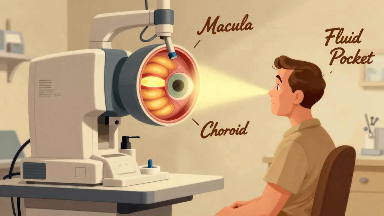
OCT is like an ultrasound for your eye, but it uses light instead of sound. It doesn’t touch your eye. You just sit in front of a machine, stare at a light, and in seconds, it builds a detailed cross-section of your retina - all 10 layers of it. This isn’t a flat photo. It’s a 3D map showing thickness, swelling, holes, or scars.
Since the late 2000s, spectral-domain OCT (SD-OCT) has been the standard. Now, newer swept-source OCT (SS-OCT) is catching up. It goes deeper, capturing the choroid - the blood-rich layer under the retina - with more clarity. It also scans faster: up to 400,000 lines per second, compared to 85,000 on older models. That means fewer blurry images from tiny eye movements.
Doctors use OCT to spot early signs of macular degeneration, diabetic swelling, glaucoma damage, and even tiny holes in the macula. In Coats disease, OCT finds fluid pockets and cholesterol crystals hidden beneath the surface - things fundus photos often miss. For diabetic retinopathy, it measures retinal thickness better than any other tool. A 2021 study showed OCT detected early fluid buildup in 92% of cases where vision was still normal.
Fundus Photography: The Classic Snapshot
Fundus photography is the oldest of the three. It’s what most people picture when they think of an eye exam - a bright flash, a colorful image of the back of the eye. Cameras like the Zeiss FF 450+ capture the optic nerve, blood vessels, and macula in full color. It’s simple, fast, and great for tracking changes over years.
But it’s limited. It only shows the surface. If there’s swelling under the retina or a thinning layer, a fundus photo won’t reveal it. That’s why it’s rarely used alone anymore. Instead, it’s the baseline. Your doctor compares this year’s photo to last year’s to see if blood vessels are leaking, if new ones are growing, or if the optic nerve is getting smaller.
In diabetic retinopathy, fundus photos are still the go-to for spotting microaneurysms and hemorrhages. But they can’t tell you if fluid is building up behind the retina. That’s where OCT steps in. In one study of 30 eyes with advanced diabetic eye disease, fundus photos caught 87% of the visible damage - but missed half the fluid causing vision loss.
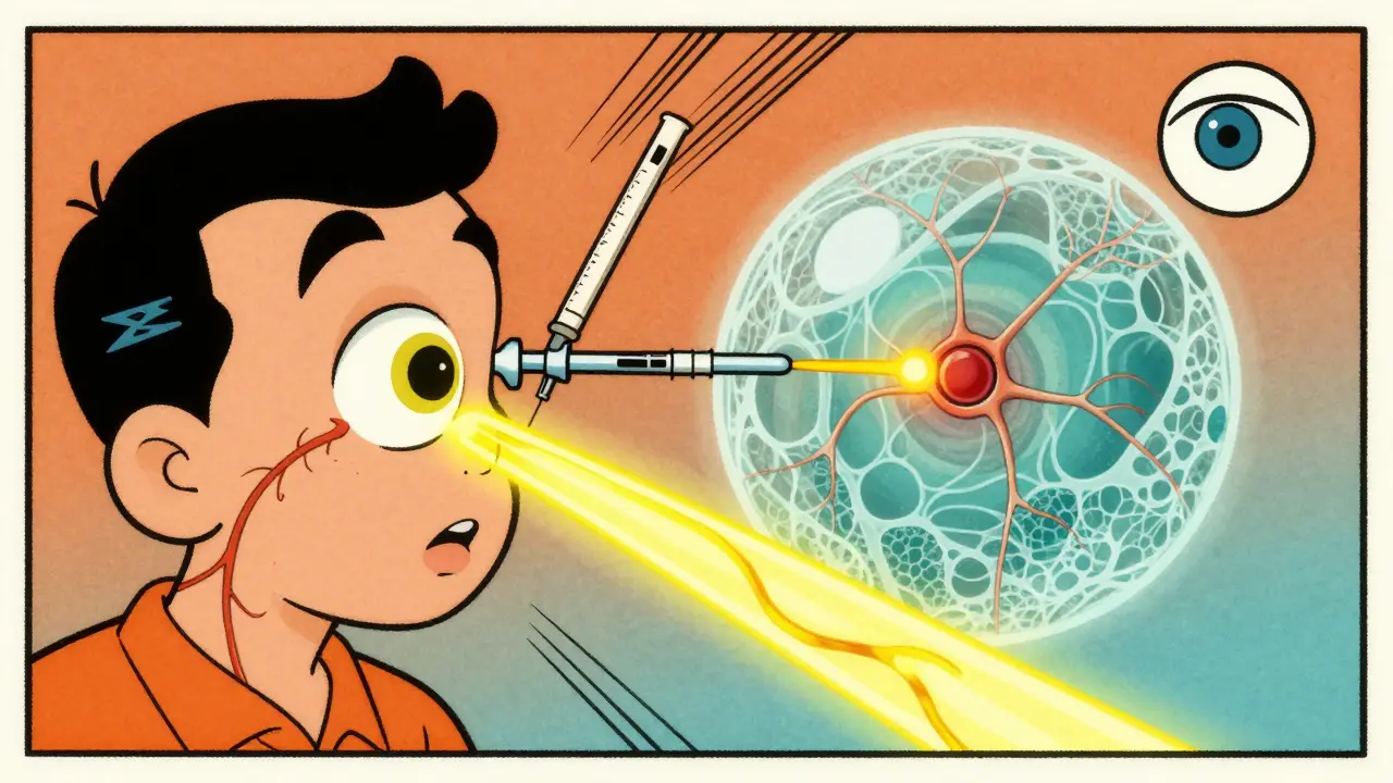
Fluorescein Angiography: The Dye That Reveals Blood Flow
Fluorescein angiography (FA) is the only one that requires an injection. A yellow dye is put into a vein in your arm. As it travels through your bloodstream, a camera takes rapid photos of your retina. The dye lights up the blood vessels, showing where they’re leaking, blocked, or growing abnormally.
This is the gold standard for seeing circulation problems. In diabetic macular edema, FA is more sensitive than OCT - it catches subtle leaks that OCT might miss. One study found FA detected leakage in 100% of cases where OCT only saw it in 79%. That’s why, even with all the new tech, FA still has a place.
But it’s not perfect. The dye can cause nausea, itching, or, rarely, allergic reactions. The whole process takes 10 to 30 minutes. And it only shows the surface vessels. It can’t tell you if the deeper layers of the retina are starving for blood.
That’s where the next tool comes in.

OCT Angiography (OCTA): Seeing Blood Flow Without the Dye
OCTA is the game-changer. It uses the same machine as OCT but analyzes tiny movements in blood cells to create detailed maps of blood flow - without any injection. You get 3D images of the superficial, middle, and deep capillary layers of the retina. No dye. No risk. No waiting.
It’s especially powerful for spotting early signs of disease. In punctate inner choroidopathy (PIC), OCTA found areas of dead blood vessels in the choroid that traditional imaging completely missed. In Coats disease, it showed abnormal vessels growing in places no other test could see. For diabetic patients, it detects tiny areas of poor blood flow before they turn into vision-threatening problems.
Studies show OCTA is better than FA at spotting neovascularization - the growth of new, fragile blood vessels - especially near the optic nerve. One 2022 study found OCTA detected 57% more of these dangerous vessels than FA in patients with severe diabetic retinopathy.
But it’s not foolproof. Motion blur from shaky eyes can ruin the image. Patients with cataracts or dry eyes sometimes get poor results. And because it doesn’t show leakage, it can’t replace FA when you need to see fluid escaping from vessels.
How Doctors Use Them Together
No single test tells the whole story. That’s why eye specialists use them as a team.
For age-related macular degeneration: OCT shows fluid buildup and drusen; OCTA reveals if new blood vessels are forming under the retina; fundus photos track pigment changes over time.
For diabetic eye disease: Fundus photos catch bleeding; FA shows leaking vessels; OCT measures swelling; OCTA finds early blood flow loss before vision drops.
For rare conditions like Coats disease or PIC: OCT finds hidden fluid and structural damage; OCTA maps abnormal vessels; FA confirms leakage patterns.
Think of it like a car diagnosis. Fundus photos are the exterior inspection. OCT is the engine scan. FA and OCTA are the fuel system check. You need all three to know if the car is running right.
What’s New and What’s Coming
Technology keeps improving. New OCTA machines now cover a wider area of the retina - up to 100 degrees - compared to the old 30-degree views. That means they can catch problems near the edge of the retina that used to be invisible.
Some systems, like the Spectralis OCTA, now use smarter software to reduce blur and improve depth. AI is starting to help too - automatically measuring vessel density, detecting early signs of disease, and flagging changes between scans.
Soon, doctors may not need to choose between FA and OCTA. New hybrid systems are being tested that combine both in one scan, giving you the leak detection of FA with the detail of OCTA - without the injection.
What to Expect During Your Scan
If you’re getting OCT or OCTA: No prep needed. You’ll sit at the machine, rest your chin, stare at a target light, and the scan takes 5-10 seconds per eye. No discomfort.
If you’re getting fluorescein angiography: You’ll get a small IV in your arm. You might feel a warm flush, a metallic taste, or mild nausea. Your skin might turn slightly yellow for a few hours. Your urine will be bright orange for a day. That’s normal. Bring sunglasses - your eyes will be light-sensitive for an hour after.
Most people find OCT and OCTA easy. FA is more involved, but it’s still safe for most. If you’ve had a bad reaction to dye before, tell your doctor. There are alternatives.
When One Test Isn’t Enough
Some patients think if one scan looks normal, they’re fine. That’s not true. A retina can look perfect on OCT but have poor blood flow only visible on OCTA. Or the blood vessels might look fine on FA, but fluid is hiding under the surface, only showing up on OCT.
That’s why doctors often order more than one. If you have diabetes, glaucoma, or macular degeneration, you’ll likely get a mix of these tests every 6 to 12 months. It’s not overkill - it’s precision.
Bottom Line: No Single Test Wins
OCT gives you structure. Fundus photos give you history. FA gives you leaks. OCTA gives you blood flow - without needles. Together, they’re the most powerful diagnostic combo in eye care today.
If you’re being monitored for an eye condition, ask your doctor: Which test is showing me what? Understanding the difference helps you see why each scan matters - and why skipping one could mean missing something important.
Is OCT safe? Does it use radiation?
Yes, OCT is completely safe. It uses harmless infrared light, not X-rays or radiation. There’s no contact with your eye, no dye, and no side effects. It’s like taking a photo with a special camera. Millions of people have had OCT scans without any issues.
Why do I need both OCT and fluorescein angiography?
OCT shows the structure - thickness, fluid, holes. Fluorescein angiography shows leaks and blood flow. One tells you what’s there; the other tells you what’s going wrong inside the vessels. For conditions like diabetic macular edema, you need both to get the full picture. One might miss what the other catches.
Can OCTA replace fluorescein angiography?
In many cases, yes - especially for spotting abnormal blood vessels or poor circulation. But OCTA can’t show fluid leaking from vessels, which FA detects clearly. For diabetic eye disease and retinal vein occlusions, FA is still the gold standard for leakage. So they’re often used together, not as replacements.
How long does each test take?
OCT and OCTA each take about 5-10 seconds per eye. Fundus photography takes 2-5 minutes. Fluorescein angiography takes 10-30 minutes total - including the injection and waiting for the dye to circulate. Most of that time is just waiting. The actual imaging is quick.
Are these tests covered by insurance?
Yes, in most cases. Medicare and private insurers cover OCT, fundus photography, and fluorescein angiography when medically necessary - like for diabetes, macular degeneration, or glaucoma. OCTA is newer, but coverage is growing fast. Always check with your provider, but if your doctor says it’s needed for diagnosis or monitoring, it’s almost always covered.
Do I need to prepare for any of these tests?
For OCT and fundus photos: no preparation. For fluorescein angiography: you don’t need to fast, but avoid caffeine or alcohol beforehand - they can make you feel more nauseous. Wear sunglasses after the test - your eyes will be sensitive to light. Bring someone to drive you home if you’re worried about blurry vision from the dilating drops.


Comments
I had my first OCT last month and honestly? It felt like sci-fi. No contact, no pain, just staring at a light and boom-your eye’s blueprint is done. My doc showed me the layers and I was like, wow, my retina has *layers*.
So calming compared to the dye stuff.
In India, we still see a lot of clinics using just fundus photos because OCT machines are expensive. But I’ve seen patients miss early diabetic changes because of it. Glad to see OCTA gaining ground-no needles, same power. Keep pushing for access!
Let’s be real-OCTA isn’t revolutionary, it’s just the latest iteration in a decades-long lineage of non-invasive retinal imaging paradigms. The real breakthrough would be multimodal integration with adaptive optics and AI-driven volumetric segmentation, which, frankly, most clinics aren’t even equipped to interpret properly. The hype around OCTA as some magic bullet ignores the fundamental limitations of motion artifact susceptibility and the irreducible diagnostic value of fluorescein leakage dynamics, which remain the physiological gold standard for capillary permeability assessment. Also, ‘no dye’ sounds nice until you realize you’re missing the functional component entirely.
As someone who’s traveled to rural clinics across the U.S. and seen how tech gaps affect care, I’ve got to say-this breakdown is exactly what we need. Not everyone has access to OCTA, but if we educate patients on why each test matters, even basic fundus + OCT can save vision. Sharing this with my community health group tonight.
I’m so glad someone finally explained this in a way that doesn’t sound like a textbook. My grandma has macular degeneration and I used to dread her appointments because I had no idea what any of the tests were for. Now I get it-OCT is like the X-ray, FA is the dye test for leaks, and OCTA is the ultrasound of blood flow. No more feeling lost in the doctor’s office. I even printed this out for her. Thank you.
Also, the part about urine turning orange? That’s wild. I told my mom and she’s now convinced the doc is poisoning her. 😅
In Nigeria, we don’t have OCTA in most hospitals. Even OCT is rare. But I’ve seen patients with diabetic retinopathy lose vision because FA was skipped-no one knew how to interpret the photos properly. This post is a gift. Hope someone translates this to pidgin and shares it in community centers.
My sister got diagnosed with PIC last year and the OCTA was the only thing that showed the dead vessel patches. The doc said without it, they’d have missed it for another year. Honestly? This tech feels like magic. I’m so glad it’s becoming more common.
OCTA is cool and all, but if your eyes are dry or you’ve got cataracts? Good luck getting a clean scan. My aunt’s results were useless because she couldn’t keep her eye still. FA still wins in those cases. Tech ain’t perfect, but it’s getting there.
octr a is lit fr no dye and u get the blood flow maps? my eye doc said its like a drone shot of your retina’s capillaries. still dont get why they do fa too tho
I used to think all eye scans were the same until I got a combo of OCT and FA for glaucoma monitoring. The FA showed a tiny leak near my optic nerve that OCT missed. Turns out, my pressure was fine but the blood flow was compromised. Scary stuff. Glad they don’t skip the old-school tests.
The article is technically accurate but dangerously oversimplified. It implies OCTA can replace FA in clinical decision-making, which is not supported by current guidelines. The absence of leakage detection in OCTA remains a critical diagnostic gap. This kind of reductionist framing risks patient harm when used as educational material in low-resource settings.
Wow, Sarah. You really do love turning a simple explainer into a 10-page peer review, don’t you?
For the rest of us who just want to know if we’re gonna need a needle or not-this post was perfect. And yes, your urine turns orange. Yes, it’s weird. Yes, you’re not dying. Now go drink water.